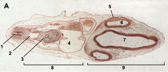
96 Hour Chick Embryo Serial Section

About Stage 13 Embryo Sections - This image is from a serial section of a 6mm CRL pig embryo with some features of the Stage 14 embryo. This embryo is approximately equal to the day 42 human embryo. Use these serial images to identify internal features and relationships that exist within the embryo at this stage. Embryo Serial Sections. 4 Day (96 hour) Chick Embryo. Extending out from the embryo is a thin delicate membrane with blood vessels at its surface. This membrane, called the allantois, expands as the embryo develops until it completely lines the shell.
Paint Tool SAI 2 free full version download OverView: Paint Tool SAI Free License Mac 1.2.5 full version developed by a very well-known company SystemX. This fantastic graphics editor application was first released on October 13, 2016. It is the best graphic editor software available in the market. Download paint tool sai full version free english. Paint Tool SAI Full Version Crack Free Download For MAC There is an extremely correct work with channels as fine. Paint Tool SAI 2018 is anything but difficult to learn yet an extraordinary straightforward client line. Paint Tools SAI is a paint program that was specially designed to facilitate manga creation. Suitable for both beginners and advanced artists, Paint Tools SAI has a very large range of tools, including superimposed layers, vector graphics, watercolor, sketch, painting, and more. Paint Tool Sai Crack 1.2.5 Free Download Full Version April 12, 2018 by fawad.getful Leave a Comment Distributed by Systemax Software Company in 2008, Paint Tool Sai cracked is a painting application and a raster graphics editor. Paint tool SAI 2.0 Crack Full Version Free Download Paint Tool Sai Crack 2.0 [Full] Free Download. Paint tool SAI crack is painting software for Microsoft Windows. It is a high-quality graphic editor with multiple instruments and effects.it helps you to apply eye-catching effects to your photos without any knowledge and experience of photo editing.
Chick Embryo 96 Hours Serial Sag Section Prepared Microscope Slide Limited Availability of Chick Embryo 96 Hours Serial Sag Section Prepared Microscope Slide EE12-3 Chick Embryo 96 Hours Serial Sag Section Prepared Microscope Slide 96 hr chick; serial sagital section A 10% discount applies if you order more than 10 of this item and 15% discount applies if you order more than 25 of this item. Triarch Incorporated offers superior prepared microscope slides. While we produce over 2300 different slides, we also make, and slides. In addition, we offer affordable quality microscopes from and for less. Use coupon code SWIFT10 for an additional 10% off our already low Swift prices. Educational digital images are also available for purchase at high resolution magnifications (10x, 25x, and 100x). CS = Cross Section: So the slide shows a thin section through the transverse plane of an organism.
LS = Longitudinal Section: So the slide shows a vertical section of the organism along the longest plane. WM = Whole Mount: So the slide shows an entire organism or structure, as indicated, is preserved on the slide CRT = Cross Section, Radial Section, and Tangential Section: So the slide shows sections of wood along the transverse, radial, and tangential planes. Sag = Sagittal Section: So the slide shows a thin section through the sagittal plane through the midline. Serial Sections = So the slide shows consecutive sections of the organism. Rep = Representative Sections (Embryology): So the slide shows one section of the organism from each typical area of study. Triarch Incorporated’s name is based on a botanical slide that illustrates three ridges of xylem found in the vascular cylinder of the Ranunculus root. Our founder, George H.
Conant, Ph.D., had three principles in mind: Accuracy, Service, and Dependability. He incorporated these into the Triarch logo based on the triarch vascular cylinder.
Another Way To Look at Serially Sectioned Frog and Chick Embryos What we show here is the first part of a project we call '4-D Embryology--Embryos in Three Dimensions and Their Changes Over Time.' We captured digital images of all the sections of serially sectioned embryos. We then used the sequence of images of an embryo to make a QuickTime digital movie. After we made movies of several serially sectioned frog and chick embryos, we started using them in our developmental biology labs.
Students like watching and using these QuickTime movies to learn about developing embryos, and we want to share these movies with other students of developmental biology. What you can see here are small 'postage-stamp' versions of these movies of serially sectioned frog and chick embryos. All show sequential cross sections going anterior-to-posterior (head-to-tail). Get Quicktime Serially Sectioned Frog Embryo Movies 4 mm 5 mm 7 mm 10 mm Serially Sectioned Chick Embryo Movies 24 hr 33 hr 33 hr 48 hr 48 hr 56 hr If you want to use these movies in your classes, you can download the full-size QuickTime movies of these embryos. To download these movies, you need to be a member of the SDB.
If you're not a member, --it really doesn't cost that much to join and it's a great organization to belong to. Anyway, e-mail me, Laurie Iten ( ), if you want to know how and where to download these movies. Why We Look At Serial Sections of Embryos When we study embryology, we need to visualize the changes that occur in developing embryos. Direct observation is the ideal method for visualizing developing embryos.
Everyone enjoys watching living embryos develop or watching time-lapse movies of developing embryos. Some embryos remain transparent throughout their development and we can see what's going on inside and outside at the same time. Most embryos are not transparent; once they become more than a few cell layers thick, we only see what's happening on the outside. To see what's going on inside, we typically cannot dissect an embryo because it's too small. We have to look at serial sections of embryos. Serial sections are where we slice an embryo as if it was a sausage. We affix each section, in order, on a microscope slide and stain the sections before we look at them with the microscope.